
Removing a mole or another skin lesion answers one of two quite different questions. Either the lesion looks suspicious and needs to be analysed, or it is harmless but catches on clothing, rubs, bleeds when you shave or simply bothers you. The approach is not the same in the two cases, and treating them as one is the commonest mistake.
That is why the visit starts with an examination, not with the removal. A doctor looks at the lesion with the naked eye and with a dermatoscope, reviews its history and only then recommends whether to remove it, how, and what should happen to the tissue afterwards. Anything removed because it raises a question is sent to a laboratory, because a microscope is the only way to get a reliable answer.
Which moles are worth removing, and when to stay closer to home
Removal during a short stay in Istanbul makes most sense for raised or bothersome moles, skin tags and other lesions that look benign, and for a lesion that a doctor wants analysed but that does not look alarming. The signs that justify a careful examination before any decision are well known:
- a change in size, shape, colour, border or thickness;
- an irregular outline or several colours within the same lesion;
- bleeding, crusting or persistent itching without an obvious reason;
- a new mole appearing in adult life, or one that looks different from all your others.
Some situations call for a different plan. If a lesion is changing quickly and looks strongly suspicious, it is often wiser to have it diagnosed and treated where you will be followed in the long term, because a confirmed melanoma may need a wider excision and further assessment by a specialist team. People with many moles, a personal or family history of melanoma, or heavily sun-damaged skin need regular surveillance of the whole skin, not the removal of a single lesion. Blood thinners, a tendency to keloid scars, an active skin infection or a very recent tan should also be discussed before planning, and a purely cosmetic removal can wait during pregnancy.
From the dermatoscope to the stitches: how removal is done
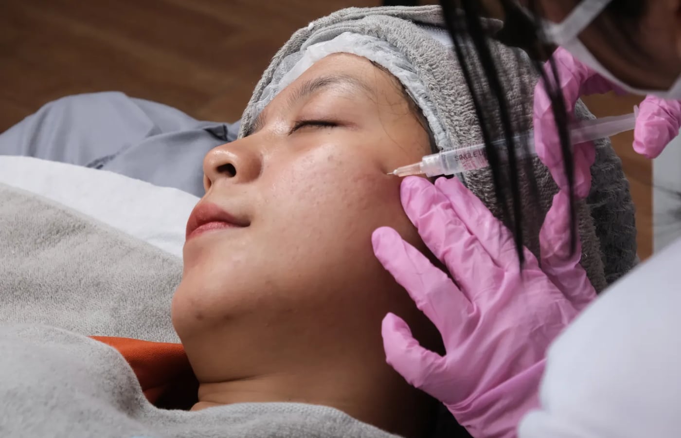
Removal is carried out under local anaesthetic, injected with a fine needle; the sting lasts a few seconds and the area then goes numb. The procedure itself usually takes around 30 minutes. The technique depends on the lesion:
- Surgical excision. The lesion is removed in full with a narrow margin of normal skin, usually as an ellipse aligned with the natural lines of the skin, and the wound is closed with stitches. This gives a complete specimen for the laboratory and is the standard approach for anything that needs to be analysed.
- Shave removal. A raised lesion that looks benign can be shaved flush with the skin. Healing is quick and stitches are not needed, but some pigment may return and the specimen is less complete.
- Punch excision. A small round blade removes small lesions, closed with one or two stitches.
- Laser or electrocautery. These techniques destroy the lesion and leave nothing to analyse. They belong only to lesions whose harmless nature is not in question after examination.
The principle is simple: a practitioner who offers to remove a lesion without examining it, or who destroys it with a laser without analysis, has removed your diagnosis along with the lesion.
Your two days in Istanbul
Day 1 – examination and removal. After arrival and transfer to your hotel, you see the doctor with an interpreter. Each lesion is examined with the dermatoscope and photographed, the options are explained and you sign a written consent. If removal is agreed, it is usually performed the same day, and you leave with a dressing.
Day 2 – wound check and flight. The wound is checked, you receive written care instructions, the plan for stitch removal and your documents, and you are taken to the airport. A small skin procedure does not usually prevent flying, and the doctor confirms that you are fit to travel. The laboratory report normally arrives a week or two later and is sent to you remotely, with an explanation.
What your written quote should cover
Esthappy organises the stay and sends a written quote listing exactly what is included. For mole removal it should cover:
- the consultation, dermatoscopic examination and photographs;
- the removal itself, local anaesthetic, stitches and dressings;
- the laboratory analysis of every specimen sent, and how the report will reach you;
- your hotel, airport and clinic transfers, and an interpreter;
- remote follow-up, including the discussion of the laboratory result.
Check that the analysis is explicitly included, and ask what is planned if the report recommends a further procedure.
Recovery, from the first day to the mature scar
First 48 hours. Keep the dressing clean and dry as instructed. A mild ache is common and settles with simple pain relief. Desk work is possible straight away.
Week 1 to 2. Stitches on the face usually come out after about a week; on the back, shoulders and limbs they stay longer, often up to two weeks, unless absorbable stitches were used. A nurse or doctor at home can remove them. Avoid stretching the area and heavy exercise while the wound is fresh, especially on the back and shoulders.
Weeks 3 to 12. The scar is pink and may feel firm. Gentle massage once the skin has closed and daily sun protection help it settle.
Months 3 to 12. The scar gradually flattens and fades. Removing a lesion always leaves a mark: on the face healing is often discreet, while on the chest, shoulders and back it is less predictable, and a widened or thickened scar is possible. Protecting the area from sun for several months is sensible.
Risks and how they are reduced
- A visible or thickened scar. Placing the incision along skin lines, closing without tension and careful aftercare reduce it; people prone to keloids are warned in advance.
- Infection or wound opening. Uncommon; clean technique and keeping the wound protected limit the risk.
- Bleeding or bruising, more likely if you take blood thinners, which is why your medicines are reviewed beforehand.
- Return of pigment after a shave removal.
- Incomplete removal or an unexpected result, which may lead the laboratory to recommend a wider excision. The risk cannot be removed, but it is managed by always sending tissue for analysis and agreeing in advance who will act on the report.
Questions to ask before booking
- Who examines the lesion, and is dermoscopy part of the consultation?
- Which technique is proposed, and why that one for this lesion?
- Where will the incision run, and how long will the scar be?
- Will every specimen be analysed, and is the analysis included?
- How and when will the result reach me, and in which language?
- Who will see me if the report calls for a wider excision? This is the question people most often forget to ask.
Back home: results, stitches and long-term checks
You go home with a written note describing what was removed, the technique, the type of stitches and the care instructions, plus photographs of the lesion taken before removal. When the laboratory report arrives, keep a copy in English together with these documents: a doctor at home who has the full report does not have to start again.
Send photographs of the wound if you have any doubt. See a local doctor for stitch removal and without delay if you notice increasing pain, spreading redness, discharge or fever. If the report recommends further treatment, the medical team explains it with you and helps you organise it, either in Istanbul or with a specialist near you. And if you have many moles or a family history of melanoma, arrange regular skin checks with a dermatologist where you live; one removal does not replace that surveillance.